Category Archives: Articles

Liquid biopsy for detection of EGFR T790M mutation in Non-Small Cell Lung Cancer (NSCLC)

Liquid biopsy for detection of EGFR T790M mutation in Non-Small Cell Lung Cancer (NSCLC)

About Non-Small Cell Lung Cancer (NSCLC)

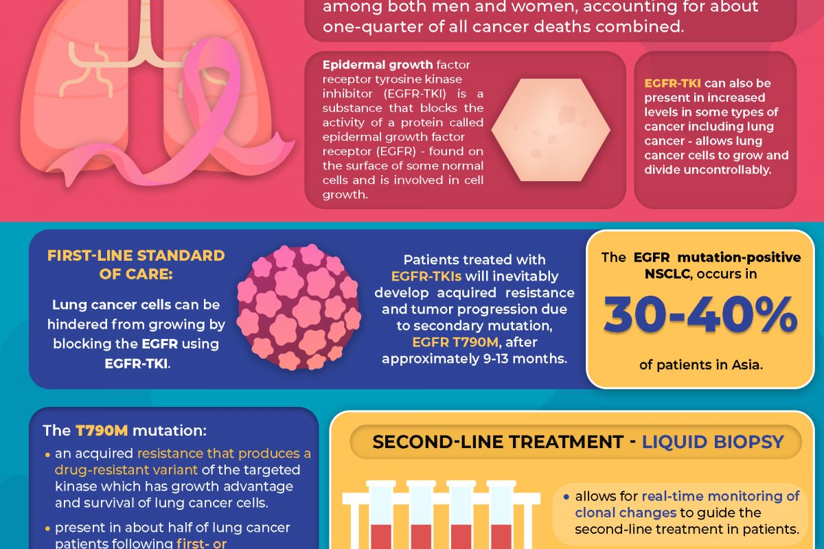
Lung cancer is the leading cause of cancer death among both men and women, accounting for about one-quarter of all cancer deaths combined.1


First-line Standard of Care

Epidermal growth factor receptor tyrosine kinase inhibitor (EGFR-TKI) is a substance that blocks the activity of a protein called epidermal growth factor receptor (EGFR), that is found on the surface of some normal cells and is involved in cell growth. This protein can also be present in increased levels in some types of cancer including lung cancer, which allows these cells to grow and divide uncontrollably.2

These cancer cells can be hindered from growing by blocking the EGFR using EGFR-TKI, making this therapy a first-line standard of care.3, 4


Acquired EGFR T790M mutation

However, patients treated with EGFR-TKIs inevitably develop acquired resistance and tumor progression due to secondary mutation, EGFR T790M, after approximately 9-13 months.3, 5, 6, 7 The EGFR mutation-positive NSCLC, occurs in 30-40% of patients in Asia.8

The T790M mutation is an acquired resistance that produces a drug-resistant variant of the targeted kinase which confers growth advantage and survival of lung cancer cells. This mutation is present in about half of lung cancer patients following first- or second- generation EGFR-TKI therapy. 9, 10, 11


Second-line Treatment

As mutational subclones may evolve over time, liquid biopsy allows for real-time monitoring of clonal changes to guide the second-line treatment in patients and to determine T790M mutation status to maximize the number of patients who are able to receive subsequent treatment with the third-generation EGFR TKI. 11, 12


Future Treatment Paradigm

Other exploratory strategies (e.g., bypass pathways, downstream signaling, histologic transformation, and others) are under development to identify patients that are suitable for molecular targeted therapy to overcome the resistance mechanisms which may change the future treatment paradigm based on preclinical and clinical studies.11


At Pantai Premier Pathology, we provide Lung Cancer Tests:

  1. EGFR Mutation Testing (Tissue)
  2. EGFR T790M mutation Liquid Biopsy Testing
  3. 170 Genes Next Generation Sequencing Testing
  4. 523 Genes Next Generation Sequencing with Tumour Mutational Burden Testing


For more information on the tests provided, please contact us at +603-42809115 (Customer Service) or email us at info@premierpathology.com.my


References

1- Key Statistics for Lung Cancer. (n.d.). American Cancer Society. Retrieved August 2, 2021, from https://www.cancer.org/cancer/lung-cancer/about/key-statistics.html

2- epidermal growth factor receptor tyrosine kinase inhibitor. (n.d.). National Cancer Institute (NIH). Retrieved August 2, 2021, from https://www.cancer.gov/publications/dictionaries/cancer-terms/def/epidermal-growth-factor-receptor-tyrosine-kinase-inhibitor

3- Bursac, D., Zarić, B., Kovačević, T., Stojšić, V., Vagionas, A., Boukovinas, I., … & Sekerus, V. (2021). Personalized Approach to Tissue and Liquid Biopsy after Failure of First-Line EGFR-TKIs: Is There an Issue When Tissue Is Not the Issue? A Case Series. Case Reports in Oncology, 14(2), 716-724.

4- Planchard, D., Boyer, M., Lee, J. S., Dechaphunkul, A., Cheema, P., Takahashi, T., … & Ohe, Y. (2018). 128O Osimertinib vs standard of care (SoC) EGFR-TKI as first-line therapy in patients (pts) with untreated EGFRm advanced NSCLC: FLAURA post-progression outcomes. Journal of Thoracic Oncology, 13(4), S72-S73.

5- Helena, A. Y., Arcila, M. E., Rekhtman, N., Sima, C. S., Zakowski, M. F., Pao, W., … & Riely, G. J. (2013). Analysis of tumor specimens at the time of acquired resistance to EGFR-TKI therapy in 155 patients with EGFR-mutant lung cancers. Clinical cancer research, 19(8), 2240-2247.

6-  Suda, K., Onozato, R., Yatabe, Y., & Mitsudomi, T. (2009). EGFR T790M mutation: a double role in lung cancer cell survival?. Journal of Thoracic Oncology, 4(1), 1-4.

7- Wu, W. S., & Chen, Y. M. (2014). Re-Treatment with EGFR-TKIs in NSCLC Patients Who Developed Acquired Resistance. Journal of personalized medicine, 4(3), 297–310. https://doi.org/10.3390/jpm4030297

8- Ellison, G., Zhu, G., Moulis, A., Dearden, S., Speake, G., & McCormack, R. (2013). EGFR mutation testing in lung cancer: a review of available methods and their use for analysis of tumour tissue and cytology samples. Journal of clinical pathology66(2), 79–89. https://doi.org/10.1136/jclinpath-2012-201194

9- Suda, K., Onozato, R., Yatabe, Y., & Mitsudomi, T. (2009). EGFR T790M mutation: a double role in lung cancer cell survival?. Journal of thoracic oncology : official publication of the International Association for the Study of Lung Cancer, 4(1), 1–4. https://doi.org/10.1097/JTO.0b013e3181913c9f

10- Choo, J. R., Tan, C. S., & Soo, R. A. (2018). Treatment of EGFR T790M-Positive Non-Small Cell Lung Cancer. Targeted oncology, 13(2), 141–156. https://doi.org/10.1007/s11523-018-0554-5

11- Liao, B. C., Griesing, S., & Yang, J. C. (2019). Second-line treatment of EGFR T790M-negative non-small cell lung cancer patients. Therapeutic advances in medical oncology, 11, 1758835919890286. https://doi.org/10.1177/1758835919890286

12- Hochmair, M. J., Buder, A., Schwab, S., Burghuber, O. C., Prosch, H., Hilbe, W., Cseh, A., Fritz, R., & Filipits, M. (2019). Liquid-Biopsy-Based Identification of EGFR T790M Mutation-Mediated Resistance to Afatinib Treatment in Patients with Advanced EGFR Mutation-Positive NSCLC, and Subsequent Response to Osimertinib. Targeted oncology, 14(1), 75–83. https://doi.org/10.1007/s11523-018-0612-z

What is COVID-19 Neutralizing Antibody Test & Why do You Need to Get Tested?

COVID-19 Neutralizing Antibody Testing

“Quantitative detections of COVID-19 Neutralizing Antibodies in human serum,

plasma or whole blood”


What is the difference between COVID-19 Neutralizing Antibody and other COVID-19 antibodies? 1, 2

Unlike other COVID-19 antibodies, COVID-19 neutralizing antibodies prevent the interaction o f S protein RBD with ACE2 cell receptor, thus, preventing the entry of virus into the host.


Application of COVID-19 Neutralizing Antibody Detection³:


Post-vaccination

To check whether your body respond to vaccination and how much of this antibody is neutralizing

antibodies; assessment of potential antibody-associated immunity

Past – Infection

To identify individuals with past exposure


Key Features³

Sensitivity: 99.45%

Specificity: >99%


  • 98% Compliance Rate when compared with FDA EUA approved ELISA test kit
  • 99.45% Compliance Rate when compared with PRNT (“gold standard”)
  • CE-marked

References:

1- Jiang, S., Zhang, X., Yang, Y., Hotez, P. J., & Du, L. (2020). Neutralizing antibodies for the treatment of

COVID-19. Nature Biomedical Engineering, 4(12), 1134-1139.

2- Neutralizing Antibody of SARS-CoV-2 Test. Packing Insert.

3- Neutralizing Antibody of SARS-CoV-2 Test. Info Sheet.

Blood Cancer & COVID-19

Blood Cancer & COVID-19

Most blood cancers, also called hematologic cancers, begin in the blood-forming tissues such as the bone marrow or in the cells of the immune system.1 Blood cancers occur when the normal blood cell development process is interrupted by uncontrolled abnormal blood cell growth, preventing functions such as fighting off infections or stopping serious bleeding.2


The three main types of blood cancers are2:

  1. Leukemia – a type of cancer that occurs in your blood and bone marrow due to rapid production of abnormal white blood cells. The high level of abnormal white blood cells are not able to fight off infections and cause impairment in the production of red blood cells and platelets in the bone marrow.


  1. Lymphoma – a type of blood cancer that affects the lymphatic system; which removes excess bodily fluids and produces immune cells. Lymphocytes are white blood cells that fight off infection. Abnormal lymphocytes turn into lymphoma cells that multiply and collect in your lymph nodes and other tissue which overtime, will impair your immune system.


  1. Myeloma – cancer of the plasma cells. Plasma cells are white blood cells that produce antibodies to fight off diseases and infections in your body. Myeloma cells prevent the normal production of antibodies causing the body’s immune system to be weakened and susceptible to infection.


Blood cancer & COVID-19

As COVID-19 emerged, patients with blood cancers are at increased risk of severe COVID-19 infection and high mortality rate due to their compromised immune system and inability to receive necessary care in time.3, 4, 5, 6 Medical practitioners have to assess whether treatment plans should begin on schedule or delayed until a certain time.5, 6  Studies have shown that patients with blood cancers are more susceptible to severe COVID-19 infection than patients with solid cancers or healthy people, likely due to the immunosuppressive nature of their disease and/or treatments. However, researchers sought to discover if immunosuppression caused patients with blood malignancies to have a decreased immune response to the COVID-19 vaccines that are now being provided around the world. 


To refer for Pantai Premier Pathology tests related to Blood Cancer, please click on the below button.

CLICK HERE


For more information on the tests provided, please contact us at +603-42809115 (Customer Service) or email us at info@premierpathology.com.my


References

1- hematologic cancer. (n.d.). National Cancer Institute (NIH). Retrieved May 4, 2021, from https://www.cancer.gov/publications/dictionaries/cancer-terms/def/hematologic-cancer

2- Blood Cancers. (n.d.). American Society of Hematology. Retrieved May 4, 2021, from https://www.hematology.org/education/patients/blood-cancers

3- Ferrara, F., Zappasodi, P., Roncoroni, E., Borlenghi, E., & Rossi, G. (2020). Impact of Covid-19 on the treatment of acute myeloid leukemia. Leukemia, 34(8), 2254-2256.

4- He, W., Chen, L., Chen, L., Yuan, G., Fang, Y., Chen, W., … & Gale, R. P. (2020). COVID-19 in persons with haematological cancers. Leukemia, 34(6), 1637-1645.

5-  Burki, T. K. (2020). Cancer guidelines during the COVID-19 pandemic. The Lancet Oncology, 21(5), 629-630.

6- American Association for Cancer Research. (2021, February). Survival Factors ID’d for Patients with Blood Cancer + COVID-19. Cancer Discovery. https://cancerdiscovery.aacrjournals.org/content/11/2/214.2


Pantai Premier Pathology COVID-19 Journey [May 2021]

Take a journey through PPP’s call of duty ensuring safety & peace of mind amidst the ongoing Pandemic. Let us not be complacent in this fight against Covid-19. Stay vigilant & stay safe!


In February 2020, Pantai Premier Pathology (PPP) received approval from the Ministry of Health as one of the 4 private labs in Malaysia to start the COVID-19 screening services. In March 2020, a total of 400 samples were run per day using the rtPCR method. These samples were received from various sites including IHH hospitals, GP clinics and corporate clients. In April 2020, PPP expanded its service offering that includes rtPCR and rapid PCR nationwide with an increase in the running capacity to 800 samples per day. A new service, RTK Antigen, was offered nationwide in May 2020, with increased running capacity to 3,500 per day. PPP has also offered support in running MOH samples when required at that time. A total YTD testing of 7.6% of the nationwide capacity at 207,396 tests was achieved in October 2020 with the launch of another service, RTK Antibody testing. In January 2021, PPP opened up another COVID-19 lab located at Pantai Hospital Ampang. After 12 months of providing COVID-19 screening service, PPP contributed to a total of 460,435 testing nationwide in February 2021. More COVID-19 screening labs were started at Gleneagles Medini, Johor Bahru and KLIA, Sepang in March 2021 with overall running capacity up to 8,000 tests per day. PPP has recorded the highest monthly total sample screened to date at 145,181 in one month as at May 2021.

Increase the Success Rate of IVF via PGS

Increase the Success Rate of IVF via PGS


In Vitro Fertilization (IVF) is an assisted reproductive technology (ART) to achieve pregnancy, where the process of fertilization is manually performed in a laboratory dish by combining extracted eggs and retrieved sperms. Once the fertilized eggs have multiplied, the embryos are then transferred to the uterus for a pregnancy to begin.1, 2, 3 This procedure is commonly conducted in couples who have difficulty in achieving pregnancy through unprotected regular intercource.2

Figure 1: The process of In Vitro Fertilisation (IVF).4


Who needs IVF treatment?

IVF can be used to treat infertility in the following cases1:

  • Unexplained infertility
  • Women with ovulation disorders; premature ovarian failure, uterine fibroids
  • Blocked or damaged fallopian tubes
  • Male factor infertility; reduced sperm count or motility
  • Individuals with genetic disorder


What is Preimplantation Genetic Screening (PGS)?

PGS is a specialised testing to screen whether cells from a cultured embryo contain any chromosomal abnormalities or balance number of chromosomes prior to implantation. PGS is highly recommended for patients with history of repeated unexplained miscarriages, recurrent IVF failures or couples with known abnormality in their chromosomal who worry passing it on to their offspring.

Common chromosomal abnormality PGS screens for are:

  1. Down Syndrome
  2. Patau Syndrome
  3. Edwards Syndrome
  4. Sex chromosomes abnormality

The process of PGS requires a small number of cells from the cultured embryos typically at day 5 (blastocyst stage) and cells are screened for normal number of chromosomes which is 46 (23 chromosomes inherited from each parents). If all chromosomes are intact with no abnormalities observed (called euploidy) then the embryo is suitable for implantation.6, 7, 10, 11, 12 Your fertility doctors will consult of the best embryos to implant in order to yield a successful pregnancy.

Figure 2: How Preimplantation Genetic Screening (PGS) Works.8


Reference

1- IVF – In Vitro Fertilization. (2019, April 24). American Pregnancy Association. https://americanpregnancy.org/getting-pregnant/infertility/in-vitro-fertilization-70966/

2- Brezina, P. R., & Zhao, Y. (2014). In Vitro Fertilization. ELSEVIER, 2014(3), 1–9. https://doi.org/10.1016/B978-0-12-801238-3.00262-2

3- Infertility FAQs. (n.d.). Division of Reproductive Health, National Center for Chronic Disease Prevention and Health Promotion. Retrieved April 8, 2021, from https://www.cdc.gov/reproductivehealth/infertility/index.htm

4- In Vitro Fertilisation. (2019, April). [Photograph]. Women’s Health Arm of Singapore Medical Group. https://smgwomenshealth.sg/news-articles/pgd-pgs-testing/

5- Harper, J. C., Geraedts, J., Borry, P., Cornel, M. C., Dondorp, W., Gianaroli, L., … & Macek, M. (2013). Current issues in medically assisted reproduction and genetics in Europe: research, clinical practice, ethics, legal issues and policy. European Journal of Human Genetics, 21(2), S1-S21.

6- De Rycke, M., & Sermon, K. (2010). Preimplantation Genetic Diagnosis. In Molecular Diagnostics (pp. 485-500). Academic Press.

7- Illumia. (2015). The STAR Trial [Brochure]. https://www.illumina.com/content/dam/illumina-marketing/documents/clinical/rgh/star-one-pager-web.pdf

8- Improving IVF outcomes with PGS. (n.d.). [Photograph]. Illumina. https://www.illumina.com/clinical/reproductive-genetic-health/preconception-fertility/pgs.html

9- Kimelman, D., Confino, R., Confino, E., Shulman, L. P., Zhang, J. X., & Pavone, M. E. (2018). Do patients who achieve pregnancy using IVF-PGS do the recommended genetic diagnostic testing in pregnancy?. Journal of assisted reproduction and genetics, 35(10), 1881–1885. https://doi.org/10.1007/s10815-018-1289-z

10- Improving IVF outcomes with PGS. (n.d.-b). Illumina. Retrieved April 9, 2021, from https://www.illumina.com/clinical/reproductive-genetic-health/preconception-fertility/pgs.html

11- Yang, Z., Liu, J., Collins, G. S., Salem, S. A., Liu, X., Lyle, S. S., Peck, A. C., Sills, E. S., & Salem, R. D. (2012). Selection of single blastocysts for fresh transfer via standard morphology assessment alone and with array CGH for good prognosis IVF patients: results from a randomized pilot study. Molecular cytogenetics, 5(1), 24. https://doi.org/10.1186/1755-8166-5-24